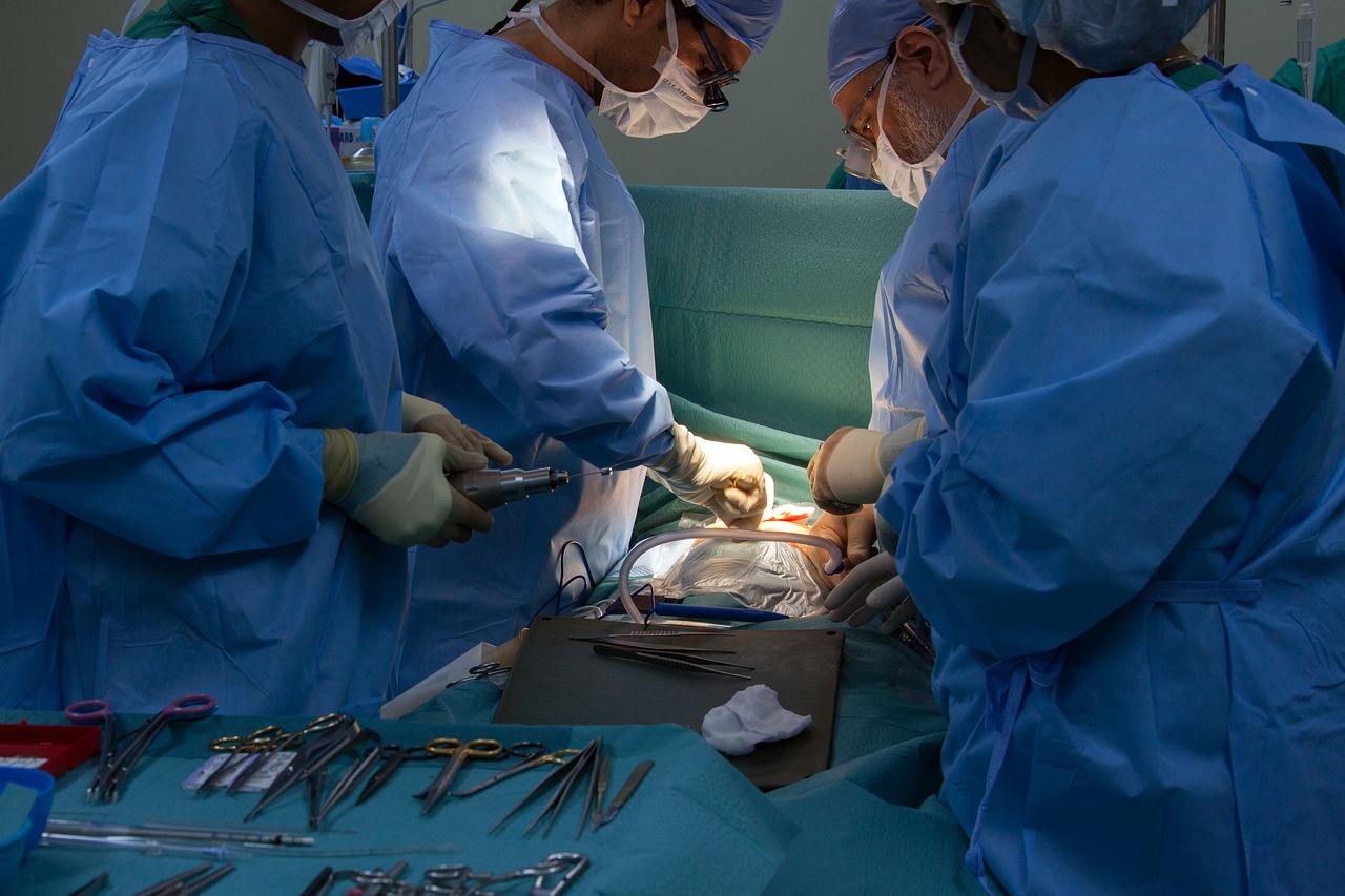
Cella Medical Solutions’ simulations can be used for pre-practice, in the interventions themselves and even materialised with 3D printers. The Murcia-based company has the capacity to generate customised models in as little as six to eight hours.
TDB keeps you informed. Follow us on Facebook, Twitter and Instagram.
Helps surgeons with its 3D
New technologies are gaining ground in operating theatres and before entering them. Doctors rely on them to know the organs or parts they are going to operate on and thus improve precision or avoid unpleasant surprises in operations.
A startup from Murcia called Cella Medical Solutions provides surgeons with medical solutions based on 3D technology, virtual and augmented reality and cloud computing. Its main activity is the creation of advanced 3D models of the patient’s organs for doctors to solve their doubts during the different phases of complex surgery.
Cella originally started out as a medical 3D printing company, but the first aspect was eventually relegated to the background.
In the process, images are taken from CT scans, MRI scans… and the anatomical structure, i.e. the organs, which can be useful for surgery, are delimited. The drawing is made by the layers that form the organ and with this, the model is constructed.
This is how the 3D simulation is created
“The process consists of taking images, such as a CT [computed tomography], PET [positron emission tomography], magnetic resonance imaging, etc., and a ‘segmentation’ is carried out. This consists of delimiting the anatomical structure, i.e. the organs, which may be of interest for surgery. This drawing process is done by the different layers that form the organ and with that, the 3D model is built,” explains Rodríguez.
This process, which is supervised at all times by a radiologist, can take longer or shorter, depending on the number of elements included in the surgery and the degree of automation, as different artificial intelligence techniques and medical imaging algorithms are used. The delineations have great ‘clinical depth’ and provide very detailed illustrations. However, the startup has the capacity to generate models in as little as six to eight hours.
“Importantly, our 3D models are tailor-made according to the doctor’s needs. It’s like a tailor-made suit,” says Cella’s technical manager. “Even if an organ is touched, sometimes we have to make complete reconstructions of areas such as the patient’s abdomen, and that is very demanding for computational purposes”. Miguel Rodriguez, CTO of Cella Medical Solutions.
Benefits for patients and doctors
Rodriguez argues that his solutions provide benefits for both patients and doctors. According to him, the former can benefit from more personalised, efficient and much more precise surgeries. In addition, intraoperative bleeding, operation time, recovery time, etc. are reduced.
As for doctors, they will be able to plan surgeries better and “go much safer”. Cella’s models not only make it possible to visualise the anatomy of patients but also to simulate certain operations and treatments. They can perform actions such as making cuts, train with certain interventions in very specific areas and practice with certain types of each organ.
Cella’s virtual models can be opened on any computer without the need to install any software or application, as they can be consulted in the web browser. “This is important because surgeons can see the 3D model at home, when travelling, on a mobile phone, on a tablet…”, says Rodríguez.
Hospitals also benefit from faster and safer interventions, which means cost savings in surgeries and savings in the postoperative period, as “this would be much quicker”, he says.
The company has the ability to produce detailed layered physical models of organs within 48 hours. / Cella Medical Solutions
From the digital to the physical world
The models created by the startup can also be easily transferred to the physical world thanks to a 3D printer. The company works not with one, but with multiple printers and, depending on the urgency, they have the possibility of materialising many organs in parallel. Thanks to this simultaneous printing with different machines, they can have each model ready within 48 hours. This is in addition to the delivery times.
With the virtual reality glasses, the surgeon can see the reconstruction floating in front of him and manipulate it. He can step inside the simulation as if he were holding it in his hand. This allows him to understand the patient’s anatomy from a more realistic perspective.
For its part, the company uses augmented reality in several ways. The main one is to assist doctors in superimposing the 3D model on a real organ. “Somehow you have to imagine it as an X-ray. With this the surgeon can see where a tumour, a vein or an important artery is, know exactly where it is because deviating a centimetre or a couple of centimetres can be a big problem,” says the CTO.
Innovations in robotic surgery
Cella also offers solutions for robotic surgeries, where doctors use incisions to insert a camera and instruments operated by a robot. The problem is that in these operations the surgeon has such an inward view of the organs that he or she can lose perspective. “We can include our 3D model inside what the surgeon sees to guide him. It’s like when you see a big screen where you see the road and below you have the GPS. We would be like ‘a GPS’ for the surgery, so to speak,” Rodriguez stresses.
The Murcian company has also developed voice commands so that surgeons do not have to use their hands for secondary areas when they are busy operating. With them, doctors can interact with the models with just their words, without the need to take their heads out of the robot’s console, go to another screen or resort to third parties. “We prevent surgeons from needing another person at their side, we help them to be autonomous,” he says.
The company has also developed voice commands so that surgeons don’t have to use their hands for secondary tasks.
Voice commands are also useful in non-robotic surgery. Another advantage of these commands is that they contribute to hygiene and prevent infections. “The surgeon touches the patient, touches the instrument, may even have blood on his hands…. The idea is to make it easier for the doctor to interact with the model without stopping him from doing what is important,” says Cella’s technical manager.
